X-rays
X-rays are electromagnetic waves with a frequency of 3x10^20 - 3x10^14 Hz and a wavelength of 10^-12 - 10^-7m
They are are part of the electromagnetic spectrum which you learnt about at AS
They are are part of the electromagnetic spectrum which you learnt about at AS
X-rays have many uses and you need to know what they are, how they are produced, how they interact with matter and how they are used
Production of X-rays
|
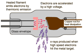
X-rays are produced when electrons strike a metal target.
The electrons are liberated from a heated filament and accelerated by a high voltage towards a metal target. The X-rays are produced when the electrons collide with the nuclei of the metal atoms |
|
Interaction of X-rays with Matter
X-rays have different forms of collisions with matter depending on the X-ray wave energies
Photoelectric effect
The photoelectric effect was introduced in AS Quantum Physics
This is a low energy phenomena collision
This is a low energy phenomena collision
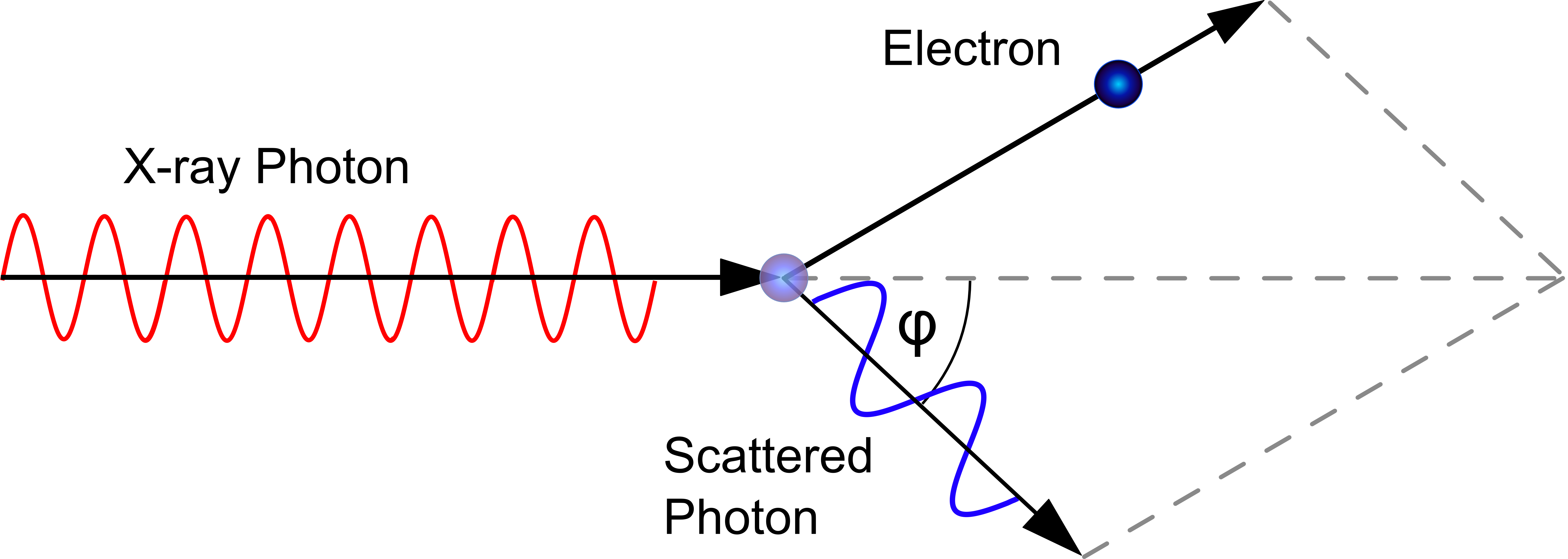
Compton Effect
This is when an X-ray photon hits an orbital electron from an atom of the absorbing material and is scattered.
Some of the photon’s energy is given to the electron.
This causes the electron to gain enough energy to leave its energy level and travels in a mirrored direction from that of the scattered photon.
The ejected electron may have any energy from 0 up to about 2/3 of the photon energy.
More energy is lost when the angle of the photon's deflection is large
This is a mid energy phenomena
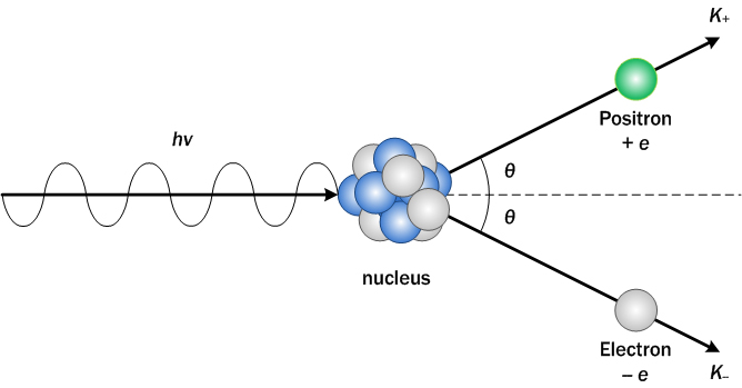
Pair Production
When an x-ray photon interacts with an atom's nucleus it splits into an electron and its antiparticle the positron provided there is enough energy for them both to exist
This is a high energy phenomena
This is a high energy phenomena
X-ray Intensity
The intensity of X-rays decreases as they travel from a POINT. They follow the inverse square law
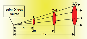
However, a collimated x-ray doesn't decrease in intensity. Collimated means all the waves travel parallel to each other
The only way to decrease a collimated x-ray's intensity is to pass it through a medium - this can be air, honey etc.
To work out the reduction in intensity the below equation is used
The only way to decrease a collimated x-ray's intensity is to pass it through a medium - this can be air, honey etc.
To work out the reduction in intensity the below equation is used
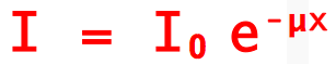
I0 = Original intensity
e = natural root - refer to C3 Exponential and Logarithms in A2 Maths
x = distance travelled through medium
μ = attenuation coefficient (in metres to the power of -1)
Attenuation occurs when an x-ray passes through a medium
The attenuation coefficient is different depending on the material
E.g.:
Vacuum = 0 m^-1
Flesh = 100 m^-1
Bone = 300 m^-1
X-rays in Medicine
|
X-rays are primarily used to view bones as they absorb more x-ray intensity than flesh and muscle
Collimated X-rays are passed through the patient onto an x-ray film. Where more x-rays are absorbed the whiter the film appears. This enables fractures and breaks in the bone to be identified as x-rays aren't absorbed as much at those points. X-rays however are low clarity 2D images as the x-rays have to pass through the whole body |

|
Improvements to X-rays
X-rays have been improved over time so to enable them to do more and be more accurate
- Ultrasensitive x-ray photographic film
- Fluorescent Intensifier screen placed behind film to provide more accurate images
- Contrast media
- Image Intensifiers
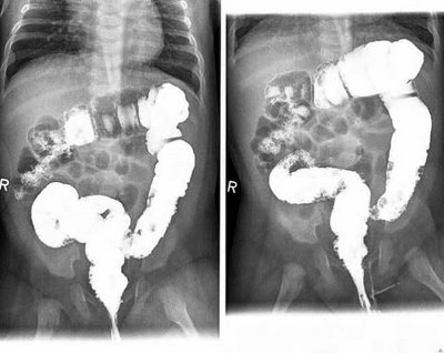
Barium Meal
|
A Barium Meal is an example of a contrast media
A barium meal is used to enable x-rays to show the difference between tissue with similar attenuation values Barium (or iodine) has a high attenuation value which means that fewer x-ray photons pass through allowing the intestines to appear. Barium is eaten and ingested coating the intestines. This enables the intestines and other soft tissue to appear in the X-rays enabling problems to be identified and a diagnosis produced |
|
Image Intensifiers
Image intensifiers turn the x-rays into visible light which are then picked up by a camera
When one x-ray photon hits the phosphur plate many visible light photons are produced. This means the intensity of the x-rays can be less
This enables real-time organ movements and high quality images
This can also lower the time the patient needs to be vulnerable to the x-rays
When one x-ray photon hits the phosphur plate many visible light photons are produced. This means the intensity of the x-rays can be less
This enables real-time organ movements and high quality images
This can also lower the time the patient needs to be vulnerable to the x-rays
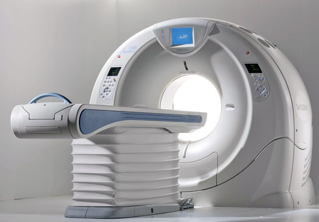
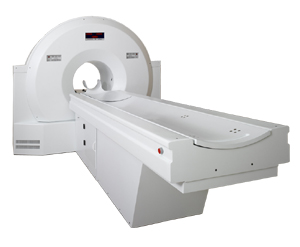
CAT Scans
|
Computerised Axial Tomography
CAT scanners have a single x-ray emitter in a column shaped chamber of x-ray detectors The emitter makes a whole revolution to produce a 3D image 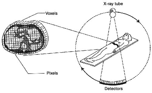
|
Advantages of CAT scans are:
|
Diagnosis Methods
Medical Tracers
- Need to be non-toxic
- Need to have a half life not too short to be ineffective or too long to be dangerous
- (Most are gamma tracers)
Technetium-99m
Technetium-99m is a gamma emitter with a half-life of 6 hours
Used to view most of the major organs
Gamma photons are emitted from the technetium-99m and its journey can be viewed enabling doctors to see how the organ is functioning
Used to view most of the major organs
Gamma photons are emitted from the technetium-99m and its journey can be viewed enabling doctors to see how the organ is functioning
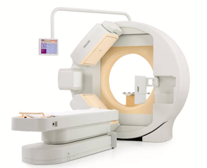
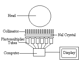
Gamma CameraA Gamma Camera is what receives the gamma rays from the medical tracer providing an image of the body
The gamma rays are emitted from inside the patient and hit the collimator, a honeycomb of lead tubes, which only gamma rays can penetrate through. These rays then reach the scintillator. This is a large sodium iodide crystal, which turns the gamma rays into visible light. These then hit the photomultiplier tubes. These create an electrical impulse that the computer will register via the below system. A gamma photon releases a photoelectron from a photocathode This then collides with a dynode and releases more electrons, which then continue to pass through several other dynodes exponentially increasing the number of electrons. These electrons then collide with an anode creating an electrical pulse which creates an image depending on the strength of the gamma rays. |
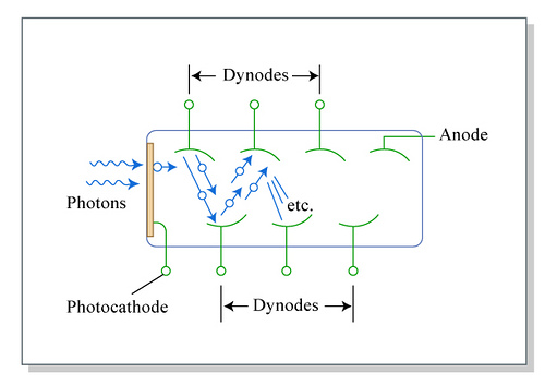
A photomultiplier tube
|
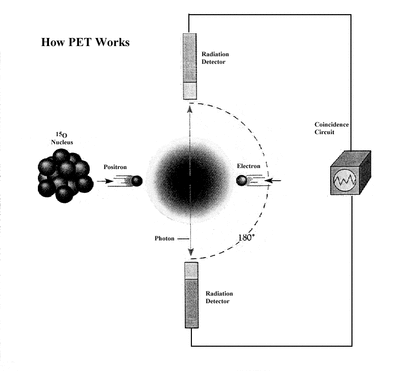
PET Scans
|
Positron Emission Tomography
PET scanners use Fluoride-18 as a tracer. It emits positrons as it decays so to become Oxygen-18 which is more stable. The positron then collides with an electron annihilating both and creating two gamma rays traveling in opposite directions to each other. Two gamma cameras then pick up the two opposite gamma rays - the difference in their arrival displays brain activity. |
|
Precession and Magnetic Resonance
|
Precession is the name given to the name for the motion that occurs at the top of an object spinning at an angle around a normal
This occurrence is caused by the torque around the normal |

|
Nuclear Spin and Precession
In a Hydrogen atom the nucleus spins creating a magnetic field with a north and south.
When Hydrogen nuclei are subjected to an external magnetic field some align themselves in the low energy state parallel to the external field. However if given enough energy they align themselves anti-parallel in the high-energy state.
When Hydrogen nuclei are subjected to an external magnetic field some align themselves in the low energy state parallel to the external field. However if given enough energy they align themselves anti-parallel in the high-energy state.
Larmor FrequencyThe precession frequency that causes the nuclei to turn anti-parallel is called the Larmor Frequency (fL) and is directly proportional to the flux density (B) of the external magnetic field. This is measured in Teslas (T). This is covered in greater detail in Electric and Magnetic Fields
The equation for Larmor is fL = 4.25x10^7 x B So, when B = 1.40 T - the larmor frequency is approximately 60MHz – a radio wave frequency |
|
Uses In Medicine
This means if the human body (10% Hydrogen) is subjected to a large magnetic field the Hydrogen molecules arrange themselves parallel to the axis of magnetism.
When a light wave of the Larmor frequency is put through the patient the Hydrogen atoms turn to the anti-parallel.
When the frequency is turned off, the hydrogen atoms turn back to the parallel position but because it is a lower energy state it must release energy. It does this by releasing Larmor frequency photons, which are detected and used to interpret tissue.
The time the nuclei take to turn back back to the parallel dictates the interpretation
Water cells take 2s and brain tissue 0.2s with cancer cells in the middle. The time it takes the hydrogen atoms to return to the parallel state is called the Relaxation Time
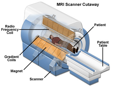
MRI Scans
|
The Magnetic Resonance Imaging scanner is made up of very big magnets
There is the main electromagnet, which gives off a magnetic field of 1.4 – 2 T, which is a constant over a 90cm central imaging section. The magnet is kept at 4.2K in liquid helium, which means that the coil has zero resistance, which means the huge current required for the huge magnetic field can be carried Covering the main magnet are gradient coils, which are accurately calibrated so as to alter the magnetic field slightly along the central imaging section. This allows a different Larmor frequency to be given off so the computer can pinpoint different tissues Also there are RF transmitter/receiver coils which give off the Larmor frequency impulses into the patient and receive the Larmor frequency waves back allowing the computer to create a detailed 3D image. Advantages
Disadvantages
|
|
Doppler Effect
The change in wavelength or frequency when there is a difference in velocity between the source and the observer
Doppler Effect in Medicine
Measuring blood flow speed
When an ultrasound transducer is held near a main artery the iron in the blood reflects the ultrasound waves.
The blood traveling towards the transducer creates a higher frequency and the blood traveling away creates a lower frequency.
The change in frequency is directly proportional to the speed of the blood and so the change in frequency can provide the speed.
Measuring Heart Beat Rate
If placed above the chest the drop in frequency levels of the blood rushing away from the heart can then allow the heart beat rate to be deduced
Ultrasound
Ultrasound is a sound wave with a frequency above 20 kHz. In medicine it usually has a range of 2 - 10 MHz
Piezoelectric Effect
This is used in Ultrasound Transducers
When a crystal or ceramic material has a p.d. applied across it, it will expand. If the material contracts a p.d. will be created.
In a crystal transducer this provides a means of converting electrical energy into sound energy
This occurs because when an alternating voltage is put through the transducer it causes the crystal to contract and expand making it vibrate. This creates an ultrasound wave to be given off.
Ultrasound Transducers
Ultrasound Transducers use the piezoelectric effect to send ultrasound waves into a patient. They than use the echoes coming back.
The echoes received are rebounds off a boundary
This boundary could be between either air and skin, liquid and tissue or tissue and bone
The distance between the boundary and the transducer can be discovered using the simple equation:
vt = d (displacement)
2
with time (t) being the time it takes to travel to the boundary and back to the transducer - therefore giving double the distance
However not all the ultrasound reflects off the boundary. Some refracts and this is due to the acoustic impedance (Z) of the new medium.
The acoustic impedance is worked out with the equation
acoustic impedance (kg/m^2s) Z = ρv density (kg/m^3) x velocity (m/s)
This is different depending on the medium the ultrasound is passing into
The intensity of the ultrasound can be discovered by using the acoustic impedances of the boundary
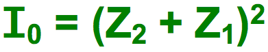
I0 = Original Intensity from transducer
Z2 = Z value of material being entered
Z1 = Z value of original Material
To discover the fraction of the intensity reflected you can use the below equation:
Z2 = Z value of material being entered
Z1 = Z value of original Material
To discover the fraction of the intensity reflected you can use the below equation:
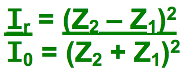
Ir = Reflected intensity
By subtracting the reflected fraction from the original intensity you can get the fraction of ultrasound that refracted
By subtracting the reflected fraction from the original intensity you can get the fraction of ultrasound that refracted
Impedance Matching
The amount reflected is important as it enables an image to be created depending on the reflection
There is a problem in medicine
Air has a Z value of 400 kg/m^2s and skin has one of 1.7 x 10^6 kg/m^2s this means that 99.9% of the ultrasound is reflected.
This would make any results completely useless.
By putting gel on the skin and transducer this removes the air - flesh boundary and so the majority of the ultrasound enters the patient
The gel has a similar Z value to flesh, this means that very little ultrasound is reflected before entering the patient giving useful results.
A-scans and B-scans
|
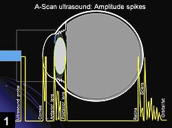
A-scans are voltage peaks on a cathode-oscilloscope - these are the points the ultrasound returns and creates a p.d using the piezoelectric effect.
This helps give the dimensions of organs. For example in an eye a simple graph would provide four peaks. The first being the pulse from the transducer, the second from the front of the eye cap, the third from the lens and the fourth from the retina. The depth of an eye can be deciphered from this information. The time taken for the depth is from the third and fourth. This is the time taken to travel from the lens to the retina and back. From this we can work out the depth, as we know the speed of ultrasound in the vitreous humour (the space in between the retina and lens), using: 2d = vt B-scans are multiple A-scans put together to create a 2D image. This is what creates uterus images
|
A-scan
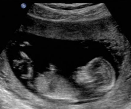
B-scan
|
Non-invasive Techniques
The above techniques are non-invasive (don't require surgery). These are important so patients are at lower risk from side effects or trauma whilst lowering costs of expensive and difficult surgery. They also speed up diagnostics.
Examples:
Examples:
- MRI
- CAT
- PET
- X-rays
- Endoscope
- Ultrasound
- Gamma Cameras
